- RESEARCH HIGHLIGHT
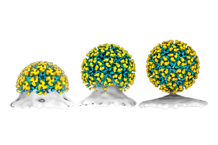
Snapshots capture self-assembly of dangerous virus

A particle of Chikungunya virus buds from a cell (artist’s illustration; spike proteins are shown in yellow; the lipid envelope in teal). Credit: D. Chmielewski et al./Nature Microbiol.
Access options
Access Nature and 54 other Nature Portfolio journals
Get Nature+, our best-value online-access subscription
$29.99 / 30 days
cancel any time
Subscribe to this journal
Receive 51 print issues and online access
$199.00 per year
only $3.90 per issue
Rent or buy this article
Prices vary by article type
from$1.95
to$39.95
Prices may be subject to local taxes which are calculated during checkout
Nature 607, 214 (2022)
doi: https://doi.org/10.1038/d41586-022-01847-0
References
Chmielewski, D., Schmid, M. F., Simmons, G., Jin, J. & Chiu, W. Nature Microbiol. https://doi.org/10.1038/s41564-022-01164-2 (2022).


