
Volume 24 Issue 7, July 2019
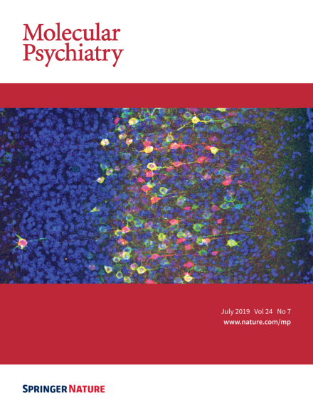
Histological image of the shRNA-CASK and a CASK deletion mutant co-transfected neurons in the somatosensory cortex of the mouse. In utero electroporation with td-Tomato (Red) and HA-tagged CASK lacking PDZ domain expression vectors was performed at E15.5 of mouse embryos and labeled pyramidal neurons in layer 2/3 of the somatosensory cortex. HA-CASK expressing neurons were visualized by the immunohistochemistry against HA tag (Green). Cortical cells were counterstained with DAPI (Blue). For more information, see the article by Mori et al. on pages 1079-1092.
Image
-
Advertisement
