Volume 14 Issue 11, November 2019
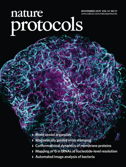
Human blood vessel organoid
Confocal image of a blood vessel organoid derived from human pluripotent stem cells, immunostained with CD31 to visualize endothelial networks (cyan) and PDGFR-b to label pericytes (magenta).
See Wimmer et al.
Image: Reiner Wimmer. Cover Design: Art Editor Erin Dewalt.