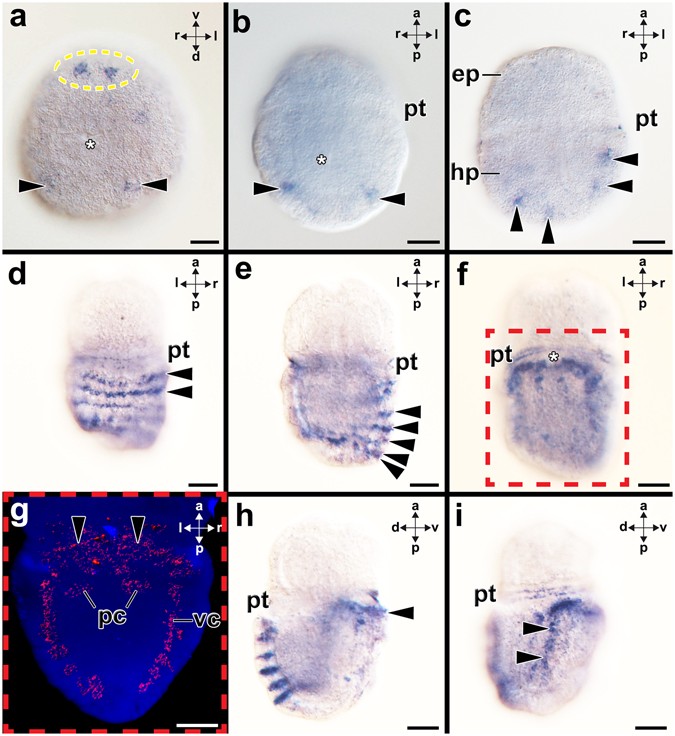
Figure 1
From: Brain regionalization genes are co-opted into shell field patterning in Mollusca

Expression of Gbx during embryonic and early larval development of the polyplacophoran Acanthochitona crinita. Dorsal (d)-ventral (v), anterior (a)-posterior (p), and left (l)-right (r) axes indicate the orientation. Asterisks mark the blastopore/mouth opening. (a) Late gastrulae (4 hpf; ventro-posterior view) expresses Gbx in cells in the ventral (encircled) and dorsal regions (arrowheads). (b-c) In further developed early trochophore larvae (12 hpf) Gbx–expressing cells (arrowheads) are located in the ventral hyposphere (hp). (d–i) Mid-stage trochophore (35 hpf). (d,e) This dorsal (d) and more ventral optical section (e) show Gbx-expressing cells (arrowheads) in seven rows of the shell fields. Note the unspecific staining in the prototroch. (f,g) Gbx is also expressed in the ventro-lateral hyposphere in the CNS, an area that is highlighted and 3-D reconstructed in (g) (red-dashed box). Gbx is expressed in the anterior pedal nerve cord (pc) and the entire visceral nerve cord (vc). Gbx is expressed in the region of the pedal nerve cord commissure (arrowheads). (h) Note Gbx-expression in the dorsal shell fields and the nervous system of the foot (arrowhead). (i) Gbx–expression in the visceral nerve cord (arrowheads). Abbreviations: ep, episphere; pt, prototroch. Scale bars: 20 µm.
