Volume 27
-
No. 12 December 2022
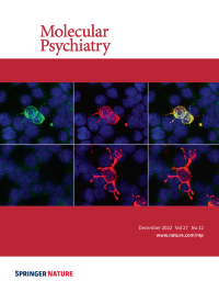
Immunocytochemical co-localization of NR1-immunized (top row) mouse plasma (1:100, green) with a commercial rabbit GluN1-AB (red) in a cell-based (HEK293T) clinical standard assay for NMDAR1-AB (Euroimmun). Double-labeled cells appear yellow (top row, right panel), documenting presence of specific circulating NMDAR1-AB in immunized mice. Plasma (1:100) of OVA-immunized mice (control immunization; bottom row) did not show specific staining for NMDAR1; GluN1/NR1, glutamate ionotropic receptor NMDA type subunit 1; OVA, ovalbumin. Counterstain with DAPI (blue) labelled cell nuclei. For more information see the article by Arinrad et al. on pages 4974–4983.
-
No. 11 November 2022
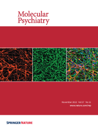
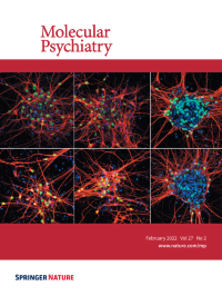
Human A9 dopaminergic neurons differentiated from induced pluripotent stem cells with the method reported in Li et al. 2022 Mol Psychiatry. Red, TH (tyrosine hydroxylase); Green, MAP2 (microtubule-associated protein 2); Blue, DNA. For more information see the article by Li et al. on pages 4407-4418.
-
No. 10 October 2022
With this global image we represent the Academy for Eating Disorders’ Truth #5: Eating disorders affect individuals of all races, ethnicities, body shapes, genders, weights, sexual orientations, and socioeconomic statuses. This piece demonstrates the global, collaborative approach that is necessary to identify both genetic and environmental causes of eating disorders with the goal of achieving better treatment and outcomes. In particular, we highlight and thank all of the people with lived experience, family members, advocates, clinicians, and researchers around the world who contribute their time, effort, and samples toward advancing eating disorders research. For more information see the article by Huckins et al. on pages 3929 – 3938.
-
No. 9 September 2022
In vivolive imaging of hypothalamic corticotropin releasing factor (CRF) neurons in the transgenicTg[crf-gal4, UAS-GFP] larval zebrafish brain with mosaic expression of the UAS-Synaptophysin-mRFP (labeling pre-synaptic terminals). For more information see the article by Wagle et al. on pages 3777 – 3793.
-
No. 8 August 2022
Shank3-deficient mice have diminished H3K4 dimethylation in PFC neurons, which is reversed by histone demethylase 1 (LSD1) inhibition. Confocal images of H3K4me2 staining in prefrontal cortex (PFC) of WT vs. Shank3 +/ΔC mice without or with the treatment of the LSD1 inhibitor GSK-LSD1 (LSD1i, 5 mg/kg, i.p., once daily for 3 days). Scale bar: 20 µm. For more information see the article by Rapanelli et al. on pages 3355 - 3366.
-
No. 7 July 2022
Single molecule fluorescentin situ hybridisation (smFISH) images of WT (left) and NLGF (right) CA1 hippocampal region. smFISH labels mRNA of Fzd1 (green), Fzd7 (yellow) and Rbfox3 (magenta) and DAPI labels nuclei (blue) show a decreased levels of expression of Fzd1 and Fzd7. For more information see the article by Palomer et al. on pages 3024-3033.
-
No. 6 June 2022
Stellate cells from the entorhinal cortex (labelled for reelin in green) project to VIP-expressing neurons in the hippocampal CA2 area (presynaptic neurons are labelled with mCherry using pseudotyped rabies virus injection in the CA2 of VIP-Cre mice. For more information see the article by Leroy et al. on pages 2879-2900.
-
No. 5 May 2022
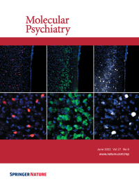
Image of the dorsal raphe nucleus stained with 5-HT, DAPI and IL1R1 shRNA expressing GFP cells. For more information see the article by Takahashi et al. on pages 2563-2579.
-
No. 4 April 2022
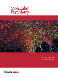
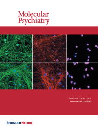
Trisomy 21 (T21) causes Down syndrome and an early-onset form of Alzheimer’s disease (AD). In this issue, Wu et al utilized human induced pluripotent stem cells (hiPSCs) along with CRISPR-Cas9 gene editing to investigate the contribution of chromosome 21 candidate genes to AD-relevant neuronal phenotypes. Shown are representative immunostaining images of neurons derived from a T21 iPSC line (bottom panels) and a T21 iPSC line which lost one copy of HSA21 (top panels). IPSC-derived neurons are immunostained for bIII-tubulin (green, left panels), neurofilament heavy chain (NEFH, red, middle panels) and POU Class 3 Homeobox 2 (POU3F2, pink, right panels). Nuclei are stained with DAPI in all images. For more information, see pages 1970-1989. Images acquired by Elizabeth Vinton.
-
No. 3 March 2022
Increased neural stem cell/progenitor apoptosis in Schizophrenia patient-derived 3D cerebral organoids. Shown are two whole-organoid images generated from a healthy adult iPSC donor (left) and from a Schizophrenia patient iPSC donor (right). Depicted is the colocalization of early SOX2+ (green) neural stem cells/progenitors and the apoptotic marker cleaved Caspase-3 (red). Cells are counterstained with DAPI (blue). We detected a substantial increase in the rate of progenitor death in Schizophrenia patient-derived cerebral organoids relative to rates observed in organoids generated from healthy control iPSC donors. In the manuscript by Notaras et al., it is shown that Pleiotrophin (PTN) but not BRN2 (POU3F2) could rescue premature progenitor death in Schizophrenia patient-derived cerebral organoids. For more information see the article by Notaras et al. on pages 1416–1434.
-
No. 2 February 2022
Human neurons with 1q21.1 deletion or duplication are associated with developmental deficits. Cortical preplate marker T-Box Brain 1 (TBR1, green, upper panel) and deep layer marker Coup-TFI Interacting Protein 2 (CTIP2, green, lower panel) were differentially expressed in iPSC derived MAP2+ neurons (red) carrying 1q21.1 deletion (centre) or duplication (right) in comparison to non-carrier controls (left). The nuclei are stained with DAPI (blue)". For more information see the article by Chapman et al. on pages 819-830.
-
No. 1 January 2022
Image showing induced pluripotent stem cells-derived excitatory and inhibitory neurons. For more information see the article by Mossink et al. on pages 1-18.