Volume 18
-
No. 12 December 2021
Reporting and reproducibility in microscopyThis month features a Focus on reporting and reproducibility in microscopy. On the cover, information regarding sample preparation, image acquisition, quality control and image analysis, collectively referred to as ‘image metadata’, is essential to ensure the interpretability, reproducibility, quality and scientific value of imaging experiments.
See Editorial
-
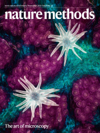
No. 11 November 2021
The art of microscopyThis image of trichomes (white), stomata (purple) and vessels (cyan) on a southern live oak leaf is the first-place winner of Nikon’s Small World Photomicrography Competition.
See Editorial
-
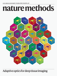
No. 10 October 2021
Adaptive optics for deep tissue imagingA compact adaptive optics module using a deformable mirror compensates for optical aberrations and enables synaptic-resolution imaging of neuronal structures in deep layers of the mouse brain.
See C. Rodríguez et al.
-
No. 9 September 2021
Cancer biology revealed with multiplexed imaging and spatial omicsSpatial omics and multiplexed imaging technologies are revealing cancer cell heterogeneity (red and purple cells) within a complex tumor microenvironment (blue, green and black cells).
See Lewis et al.
-
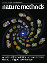
No. 8 August 2021
An atlas of transcription factor expression during C. elegans developmentA single-cell atlas of transcription factors mapped onto C. elegans cell lineage information reveals mechanisms of fate patterning and regulation during embryogenesis.
SeeMa et al.
-
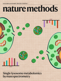
No. 7 July 2021
Single-lysosome metabolomics by mass spectrometrySingle-lysosome mass spectrometry measures the metabolomic heterogeneity of individual enlarged lysosomes.
See Zhu et al.
-
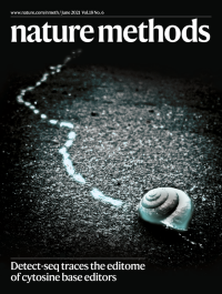
No. 6 June 2021
Detect-seq traces the editome of cytosine base editorsJust as the path of a snail can be traced by the trail of slime it leaves behind, Detect-seq traces the editing-intermediate deoxyuridine to reveal the editome, including different types of off-target mutations induced by cytosine base editors.
See Lei et al.
-
No. 5 May 2021
PDB 50th anniversary: celebrating the future of structural biologyIn honor of the 50th anniversary of the Protein Data Bank (PDB), we and our colleagues at Nature Structural & Molecular Biology present a joint special focus issue to celebrate both the past and the future of structural biology.
See Editorial
-
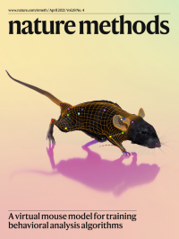
No. 4 April 2021
A virtual mouse model for training behavioral analysis algorithmsA 3D virtual mouse model was generated by adding features such as fur and whiskers to a mesh model of the body. After annotation with landmarks and generation of realistic videos of behavior, the virtual mouse model can be used to train behavioral analysis algorithms.
See Bolaños et al.
-
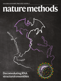
No. 3 March 2021
Deconvoluting RNA structural ensemblesDRACO is an algorithm for the deconvolution of coexisting alternative RNA conformations from structure probing experiments. When applied to the SARS-CoV-2 genome, DRACO revealed the presence of structurally dynamic regulatory regions, including the frameshifting element and the 3′ UTR.
See Morandi et al.
-
No. 2 February 2021
A high-efficiency exosome isolation systemEXODUS is an ultrafiltration strategy for purifying exosomes from biological fluids with high efficiency. Periodic negative pressure oscillations across a nanoporous membrane filter allow impurities and liquids to pass through while trapping the exosomes in the central chamber.
See Chen et al.
-
No. 1 January 2021
Method of the Year: spatially resolved transcriptomicsOur choice for the 2020 Method of the Year is spatially resolved transcriptomics. The cover depicts an example of data generated by spatially resolved transcriptomics technology. Middle layer: H&E-stained small intestine section. Bottom layer: mRNA capture platform (for example, barcoded microarray or beads). Top layer: RNA-seq data from the small intestine section.
See Editorial